3D Printed Tungsten Collimator
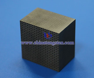
1. What’s 3D Printed Tungsten Collimator?
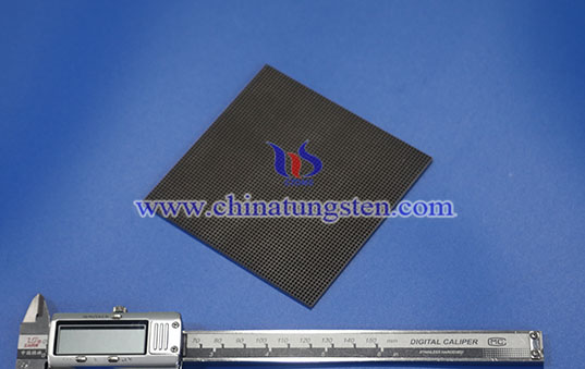
3D printed tungsten collimator is radiation beam control component manufactured through Additive Manufacturing (AM) technology using pure tungsten or tungsten alloy and formed layer by layer. Tungsten’s high atomic number (Z=74) and high density enable effective absorption and attenuation of X-rays, γ-rays, and other radiation, while channels, grids, and slits restrict beam divergence and reduce scattered radiation. Compared with conventional machining or assembly, 3D printing better suits thin-wall, micro-hole, and complex integrated structures for CT, X-ray imaging, nuclear medicine, and research detection equipment.
2. Features of CTIA’s 3D Printed Tungsten Collimator
(1) Complex Integrated Structures: Manufacturing honeycomb, grid, micro-hole, and curved-channel structures that are difficult to produce through conventional machining, reducing structural joints and assembly errors.
(2) High Shielding Efficiency: Pure tungsten and high-tungsten-content alloys provide effective radiation attenuation, with pure tungsten density of approximately 19.25 g/cm³, suitable for structures requiring high shielding efficiency.
(3) Fine Structural Features: Small apertures, thin walls, and high-density arrays, with feature dimensions optimized according to equipment and process requirements.
(4) Flexible Beam Control: Customized channels and shielding regions based on radiation energy, beam direction, field of view, and detector structure for targeted beam control.
(5) Controlled Post-Processing Precision: Grinding and precision machining after 3D printing improve dimensional accuracy of critical mating surfaces, channels, and mounting locations.
(6) High Material Utilization: Near-net-shape manufacturing and structural optimization reduce material waste, supporting complex, small-batch, and customized collimator production.
3. Applications of CTIA’s 3D Printed Tungsten Collimator
(1) CT and X-ray Imaging: Used for detector-front collimation and scatter radiation control to reduce unwanted radiation entering detectors and improve image quality.
(2) Nuclear Medicine Imaging: Used in pinhole, parallel-hole, and other collimation structures in Single Photon Emission Computed Tomography (SPECT) and other equipment to control γ-ray incidence direction.
(3) Radiotherapy: Used for high-energy X-ray beam shaping and localized shielding to help form radiation fields according to treatment plans.
(4) Industrial Non-Destructive Testing: Used in X-ray and γ-ray radiography equipment to restrict beam coverage and reduce scattered radiation.
(5) Research and Detection Equipment: Used in radiation detectors, experimental radiation fields, and high-energy physics equipment for beam control and anti-scatter structures.
4. Specifications of CTIA’s 3D Printed Tungsten Collimator
(1) Materials: Pure tungsten (W), W-Ni-Fe tungsten alloy
(2) Density: Pure tungsten ~19.25 g/cm³; tungsten alloy 16.7–18.8 g/cm³
(3) Structures: Parallel-hole, pinhole, slit, honeycomb, micro-hole arrays; customizable
(4) Dimensions: Overall dimensions, aperture, pitch, wall thickness, customizable
CTIA GROUP has nearly 30 years of experience in tungsten and tungsten alloy manufacturing, with expertise in precision machining of high-density tungsten shielding components. CTIA combines 3D printing with precision machining to provide 3D printed tungsten collimators with customized materials, channel structures, dimensions, and machining precision for CT, nuclear medicine, radiotherapy, industrial inspection, and research equipment, supporting complex radiation beam control and precision assembly.
For any inquiry, please contact tungsten alloy manufacturer: CTIA GROUP
Email: sales@chinatungsten.com
Tel: 0086 592 5129696 / 0086 592 5129595
Website: tungsten-alloy.com
WeChat:

Related Products: